


Optical Microscopy Application: Brightfield Illumination
作者: Stephan Briggs
Brightfield illumination, which yields dark objects on a bright background, is the simplest technique for optical microscopy. In brightfield illumination, the light source is positioned below the sample. Light then propagates through the sample, and is observed by the objective lens and sensor, which are positioned above the sample. The darker the sample, the denser the specimen, as denser samples absorb more light. The simplicity of brightfield illumination is the main reason this technique is so popular in optical microscopy.
Image Appearance
A typical brightfield illumination image has a dark sample with a white background. The darker the regions on a sample, the more absorption of light that has occurred. For example, plant cells would appear darkest at the nucleus and central region where cellular matter is most dense, and lighter in the cytoplasm void of ribosomes, the endoplasmic reticulum, and other intracellular components. Animal cells are more difficult to image with this technique without staining of the sample, which ultimately kills live cells.
Figure 1: Brightfield Illumination Image of Tissue Paper
Technical Details
There are four key components in the light path for brightfield illumination.
- Light Source: trans-illumination below sample that propagates through to condenser and objective lens. Typically a broadband source such as a quartz halogen bulb.
- Condenser Lens: collects trans-illuminated light and focuses to sample.
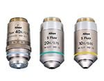
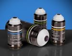
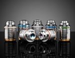
- Objective Lens: collects light which propagates through sample and enhances details by a factor of magnification.
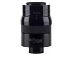
- Eyepiece/Camera: views or records the image.
There are some limitations to the brightfield illumination technique, which include very low contrast for cellular or biological samples, low optical resolution due to limitations of light, and the requirement for stained samples prior to imaging or viewing. However, simplicity of the brightfield technique is a huge benefit when first imaging an unknown sample. Additional optical microscopy applications include darkfield illumination, phase contrast, fluorescence, and differential interference contrast.
或查看各区域电话
报价工具
只需输入商品编号
Copyright 2023, 爱特蒙特光学(深圳)有限公司。— 广东省深圳市龙华工业东路利金城科技工业园3栋5楼 518109 - 粤ICP备2021068591号